 |
| Pipetting during DNA extraction. |
 |
| Preparing a perfectly even gel for electrophoresis at sea. |
 |
| The buoy marking the center of station ALOHA. |
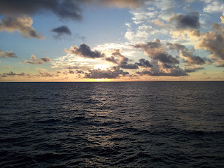 |
| Subtropical open ocean sunrise at 06.00. |
 |
| The final size of the cups after being lowered down to 4790 m. |
 |
| Happy upcoming birthday, my son. This one is for you! |
 |
| A selfie with the flow cytometer. |
 |
| Be mindful of your surroundings when working in the rad-van. |
I hope the pictures mostly speak for themselves. I was thinking that that should be it for this post but I just feel like I have to write a few lines about the deep cast and the flow cytometer. I was too excited about it, not to.
I can just mention that we did have a "biodiversity-day" (first two pictures) with Grieg Steward and Craig Nelson, who by the way were awesome to work with. This day comprised of DNA-extraction, PCR (polymerase chain reaction) and gel electrophoresis. This is what I do, so naturally it was my moment to shine!
Next up was the flow cytometer with Ken Dogget. This was an instrument/method I was most excited about on the entire cruise. It truly is an ingenious and complex instrument but the application still is fairly simple and unless you're a flow cytometer technician, most of the complicated work included is interpreting the data plots that the instrument gives.
So what the instrument actually does is counting and sorting cells in a water sample. Thats right individuals cells! Im far from an expert on this instrument after only working with it for a day but to simplify how it works, it uses laminar flow to dispense the sample into a so called interrogation zone (where the sample is read by the instrument). Here it shoots a laser beam at the sample which will scatter and fluoress when it hit cells. The angle and light is dependent on cell size and incorporated pigments (you can also dye your sample). This light signal is then picked up by detectors (they do have a fancy name which I can't remember now) which will eventually give a "pixel" in a scatter plot on a computer. Now it is a matter of interpretation and hopefully (and most often in these waters) you can see distinct clusters of cells (one pixel being one cell) and can then make out size ranges and/or pigment intensities of species or populations of microbes in the water, by visualizing them in different light (e.g. chlorophyll in red light).

So that was counting cells, but you can also sort cells, meaning that you can make the instrument put all cells (within a range) of your choosing in a tube, separating up to four species or populations at the same time, from the same sample water. This is achieved by use of electricity and polarization. So the tiny flow of your sample water going through the interrogation zone is made to vibrate, by adding a low voltage to the nozzle. This causes the water to make tiny waves which eventually break off into droplets. The trick here is to calculate and instruct the instrument as to how long it takes for a "laser-read" cell to end up in the first droplet formed down the flow. This droplet will then be charged and easily sorted. The calibration of this operation is mainly done using tiny cameras inside the instrument.
It might sound like this is a process which would take a while, and to be frank, yes it does, BUT the only reason for that is that we have such large quantities of microbes in our waters. The instrument is capable of counting and sorting at a staggering rate of 20 000 cells/sec!
Finally, I unfortunately lost one cup during the deep cast. I had made three, one for each of my kids back home. Apparently there's another deep cast going down later (2000 m) which should be enough to shrink the cups. So I made another cup. Here it is. Hopefully I won't lose it.


 So that was counting cells, but you can also sort cells, meaning that you can make the instrument put all cells (within a range) of your choosing in a tube, separating up to four species or populations at the same time, from the same sample water. This is achieved by use of electricity and polarization. So the tiny flow of your sample water going through the interrogation zone is made to vibrate, by adding a low voltage to the nozzle. This causes the water to make tiny waves which eventually break off into droplets. The trick here is to calculate and instruct the instrument as to how long it takes for a "laser-read" cell to end up in the first droplet formed down the flow. This droplet will then be charged and easily sorted. The calibration of this operation is mainly done using tiny cameras inside the instrument.
Inga kommentarer:
Skicka en kommentar